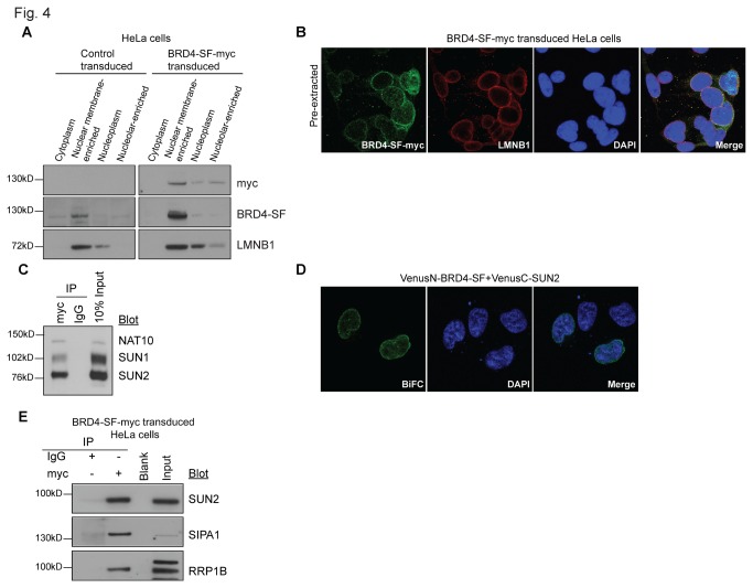
Figure 4. Epitope-tagged BRD4-SF localizes at nuclear membrane and interacts with SUN2.
(A) Cellular fractionation of HeLa cells stably expressing myc-tagged BRD4-SF using Lamond lab protocol followed by western blot analysis. Control cells were transduced with an empty vector. (B) Confocal microscopy showing co-immunofluorescence of HeLa cells ectopically expressing myc-tagged BRD4-SF (green) after co-staining with the nuclear envelope marker lamin B1 (LMNB1; red) and double-stranded DNA (DAPI; blue). (C) Immunoprecipitation of HeLa cells stably expressing myc-tagged BRD4-SF using modified formaldehyde cross-linking protocol and anti-myc antibody for immunoprecipitation followed by western blot analysis. (D) BiFC analysis of BRD4-SF and SUN2 in HeLa cells followed by confocal microscopy. Green: fluorescence complementation, blue; DAPI: double-stranded DNA. (E) Immunoprecipitation from nuclear membrane-enriched fraction of HeLa cells ectopically expressing myc-tagged BRD4-SF using anti-myc antibody for immunoprecipitation followed by western blot analysis. All pictures were taken at 63x magnification using confocal microscopy.

