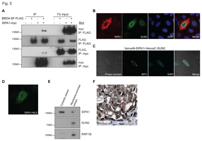
Figure 5. BRD4-SF interacts with SIPA1.
(A) Interaction of epitope-tagged BRD4-SF and SIPA1 by reciprocal Co-IP in HEK293 cells using antibodies against the epitope tags, followed by western blot analysis. (B) Co-immunofluorescence of epitope-tagged SIPA1 (red) and SUN2 (green) in HeLa cells. Blue; DAPI: double-stranded DNA. (C) BiFC analysis of SIPA1 and SUN2 in HeLa cells followed by confocal microscopy. Green: fluorescence complementation, blue; DAPI: double-stranded DNA. (D) Immunofluorescence of a SIPA1 construct that has an SV40 nuclear localization signal (green) in HeLa cells followed by confocal microscopy. (E) Western blot analysis performed on the cytoplasmic fraction of HeLa cells isolated using Lamond lab cellular fractionation protocol after high speed centrifugation at 100,000 x g. Supernatant: cytosolic extract, pellet: membrane-enriched extract. (F) Immunohistochemical staining of breast cancer TMAs from the Human Protein Atlas for SIPA1 protein. All confocal microscopy pictures were taken at 63x magnification.

