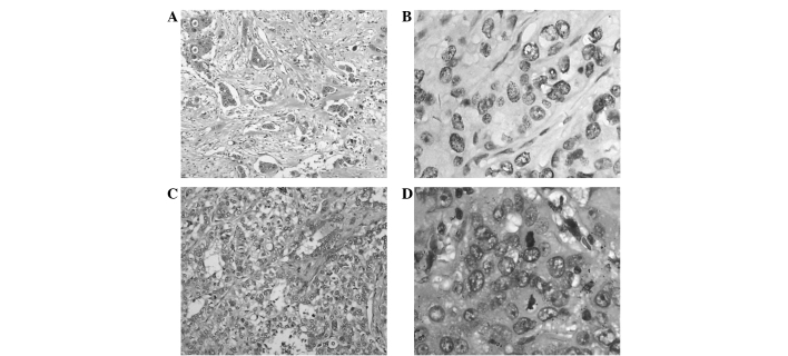
Figure 1.
Bright field pathology images. (A) Primary lung tumor (HE staining; magnification, ×100). Tumor solid growth with evident differentiation. (B) Metastasis (IHC staining with TTF1-antibody showing positive tumor with high magnification (x400). The tumor demonstrated an increased size with evident differentiation. Mitosis was observed. (C) Metastasis (HE staining; magnification, ×100). Invasive growth of tumor cells in fibrous tissue. (D) Metastasis (HE staining; magnification, ×400). Tumor cells with an abundance of cytoplasm. Mitosis were observed. HE, hematoxylin and eosin; IHC, immunohistochemistry.

