Figure 1.

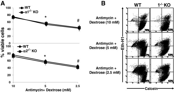
Effect of metabolic stress on the viability of MPT cells from α1-/- and α2-/- KO mice. A: MPT cells from α1-/- mice (upper panel) or α2-/- KO mice (lower panel), or their WT controls, were subjected to graded metabolic stress by incubation in the presence of antimycin (2 μM) and varying concentrations of dextrose. After 16–18 hrs, the percentage of viable cells remaining was determined by flow cytometry (n = 5 for both experiments). * p < 0.02, 5 mM dextrose vs. 10 mM dextrose; # p < 0.02, 2.5 mM dextrose vs. 5 mM dextrose. B: Representative flow cytometric analysis of MPT cells from α1-/- mice (right panels) and their WT control (left panels) incubated in antimycin (2 μM) plus varying concentrations of dextrose. Cell viability was quantified by staining cells with calcein AM (x-axis) and ethidium homodimer-1 (Eth-H1) (y-axis). For each condition, 10,000 cells were analyzed, and the percentage of viable cells was calculated by dividing the number of calcein-positive and Eth-H1-negative cells by the total number of cells counted.
