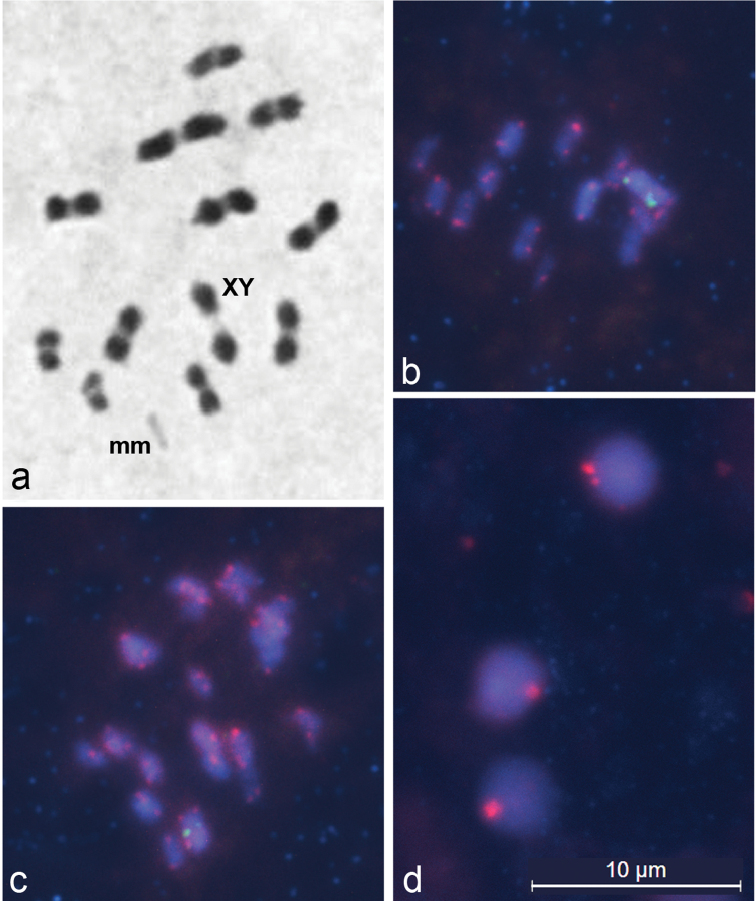
Figures 1.
Meiotic chromosomes of Lethocerus patruelis subjected to standard staining (a) and FISH (b–d). a metaphase I showing n = 11AA + mm + XY; b–d representative FISH images of metaphase I chromosomes (b, c) and spermatids (d) hybridized with probes against 18S rDNA and telomeres, showing ribosomal clusters (green) on X and Y chromosomes (b, c), and TTAGG repeats (red) located at the ends of chromosomes (b, c) and clustered at the periphery of spermatid nuclei (d).

