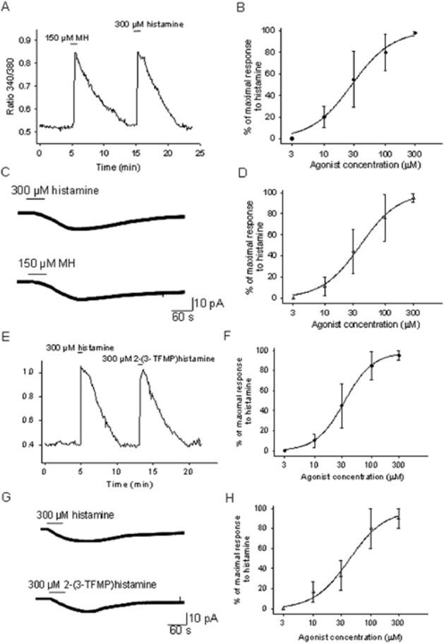
Figure 2.

Methylhistaprodifen (MH) and 2-(3-TFMP) histamine are full agonists at the H1 receptors expressed by PO/AH neurons. (A) [Ca]i responses to histamine (300 μM) and MH (150 μM). The trace represents the average the response from six cultured PO/AH neurons. (B) Concentration–response curve for the activation of [Ca]i responses by MH normalized to the response to histamine (300 μM). Each point represents the average of data collected from 30 different PO/AH neurons. The data were fitted with a Hill function. The fit yielded an EC50 of 28 μM. (C) Inward current activated by histamine (300 μM) and by MH (150 μM) recorded in a cultured PO/AH neuron. The holding potential was −50 mV. (D) Concentration–response curve for the inward currents activated by MH. The values were normalized to the maximal inward current activated by histamine (300 μM) in the same neuron. Each point represents the average of data collected from eight different PO/AH neurons. The data were fitted with a Hill function. The fit yielded an EC50 of 32 μM. (E) [Ca]i responses to histamine (300 μM) and 2-(3-TFMP) histamine (300 μM). The trace represents the average of the responses from 5 cultured PO/AH neurons. (F) Concentration–response curve for the activation of [Ca]i responses by 2-(3-TFMP) histamine normalized to the response to histamine (300 μM). Each point represents the average of data collected from 28 PO/AH neurons. The data were fitted with a Hill function. The fit yielded an EC50 of 36 μM. (G) Inward current activated by histamine (300 μM) and by 2-(3-TFMP) histamine (300 μM) recorded in a cultured PO/AH neuron. The holding potential was −50 mV. (H) Dose–response curve for the inward currents activated by 2-(3-TFMP) histamine. The values were normalized to the maximal inward current activated by histamine (300 μM) in the same neuron. Each point represents the average of data collected from 6 different PO/AH neurons. The data were fitted with a Hill function. The fit yielded an EC50 of 41 μM. (A–H) TTX (1 μM), CNQX (20 μM), AP-5 (50 μM), and bicuculline (20 μM) were present in the extracellular solution.
