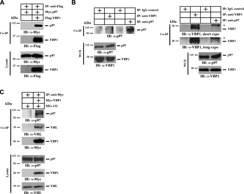
Figure 2.
VBP1 interacts with p97. A) HEK293T cells were transfected to express 3xFlag-VBP1 and Myc-p97. Cells were lysed 24 h post-transfection, and 10% of cell lysates were saved as input. VBP1 was immunoprecipitated with an anti-Flag antibody, and p97 was examined using an anti-Myc antibody. Co-IP, coimmunoprecipitation. B) Whole-cell extracts (WCE) of HEK293T cells were prepared, and immunoprecipitation was carried out using either anti-VBP1 or anti-p97 antibody. IgG was used as a negative control. Asterisk indicates Ig light chain. C) Both parental HEK293T cells and those transfected with Myc-VBP1 were treated with 5 μM MG-132 for 12 h to block proteasome-mediated protein degradation. Cell lysates were then subjected to immunoprecipitation using an anti-Myc antibody. Endogenous p97 and VHL were analyzed in the VBP1 immunocomplex.

