
Figure 4.
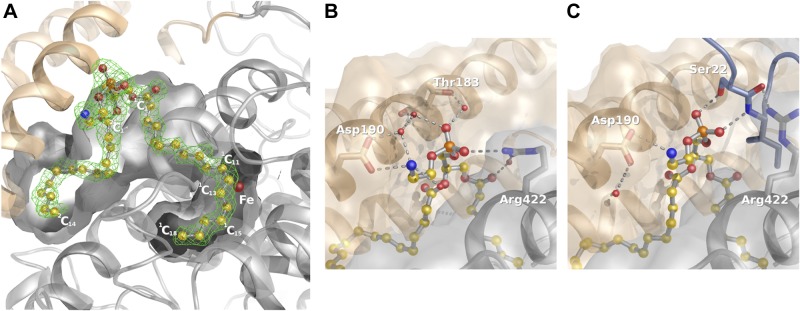
Phospholipid binding to Pa_LOX. A) Section of the binding-pocket showing a 1.75 Å resolution omit electron density map at 2.5 σ (green) of the molecule found in the substrate binding pocket of Pa_LOX. The density, which allows positioning of all the nonhydrogen atoms, corresponds to a PE molecule (balls and sticks). B) The presence of 2 ionic bonds between Pa_LOX and the PE head in the crystal form determined at 1.75 Å resolution suggests specificity toward small polar or charged head phospholipids. C) However, interactions of the PE head are altered in the crystal form determined at 2.0 Å resolution because of the presence of the N-terminal tail of a neighboring molecule (bluish gray).