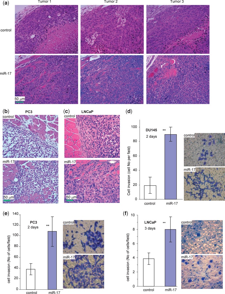
Figure 5.
miR-17 affects tumor cell invasion. (a) The H&E stained tumor sections were examined under a light microscope. Evidence of invasive tissues could be seen in the miR-17 DU145 tumors, but it was absent in the control tumors. (b and c) Evidence of invasive tissues could also be seen in the miR-17 PC3 tumors (b) and miR-17 LNCaP tumors (c), but not in the control tumors. (d–f) Cell invasion was assayed in miR-17- and vector-transfected DU145 (d), PC3 (e) and LNCaP (f) cells using Matrigel-coated transwell chambers. The miR-17 cells were significantly more invasive than the control cells (left, **P < 0.01, n = 5). Typical photos are shown (right).

