Figure 8.
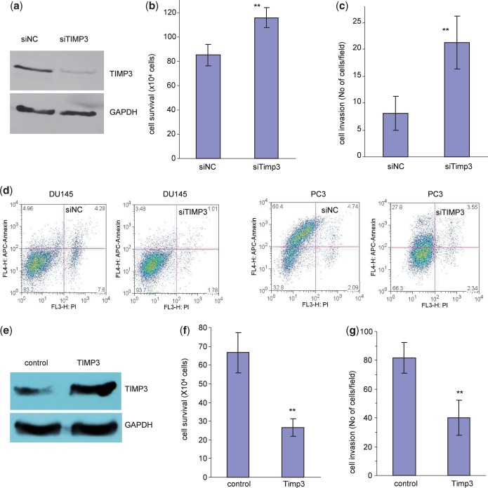
Confirmation of TIMP3 in mediating miR-17 functions. (a) Cell lysate prepared from DU145 cells transiently transfected with a control oligo or siRNA oligo targeting TIMP3 was subject to western blot analysis probed with anti-TIMP3 antibody to confirm silencing of TIMP3 expression by siRNA transfection. (b) DU145 cells transiently transfected with the TIMP3 siRNA or the control oligo were grown on 12-well tissue culture dishes in serum-free conditions for survival assay. Transfection with siRNA enhanced cell survival. **P < 0.011, n = 5. (c) DU145 cells transiently transfected with the TIMP3 siRNA or the control oligo were placed on matrigel coated transwell membranes for invasion assay. Transfection with siRNA enhanced cell invasion. **P < 0.001, n = 5. (d) DU145 and PC3 cells transiently transfected with the TIMP3 siRNA or the control oligo were examined for cell apoptosis. Transfection with siTIMP3 enhanced apoptosis of both types of cells. **P < 0.001, n = 3. (e) Cell lysates prepared from DU145 cells stably transfected with miR-17 and transiently transfected with TIMP3 expression construct or a control vector were subject to western blot analysis probed with anti-TIMP3 antibody. TIMP3 expression was increased by TIMP3 transfection. (f) The cells were subjected to cell survival assays. Transfection with TIMP3 reversed the effect of miR-17 resulting in decreased cell invasion. **P < 0.01, n = 10. (g) The cells were also subjected to cell invasion assays. Transfection with TIMP3 reversed the effect of miR-17 resulting in decreased cell invasion. **P < 0.01, n = 5.

