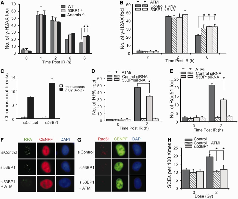
Figure 1.
53BP1 promotes DSB repair by HR in G2. (A) Wild-type (WT), 53BP1−/− and Artemis−/− MEFs were exposed to 3 Gy IR and γH2AX foci enumerated to 8 h post-IR. G2 cells were identified by CENPF staining (11). Aphidicolin was added before IR to prevent S phase cells progressing into G2 during analysis in all experiments. This also enhanced the distinction between S and G2 cells, as aphidicolin induces pan nuclear γH2AX formation in S phase cells. Control experiments verified that this does not affect HR in G2 (4,11). (B) γH2AX foci enumeration in A549 cells with or without siRNA 53BP1 following 3 Gy IR. ATMi was added as indicated. (C–H) Chromosomal breaks (C), RPA foci (D), RAD51 foci (E) and SCEs generated from G2 cells (H). RPA and RAD51 foci were enumerated at 2 h post 3 Gy IR. Chromosomal breaks and SCEs were scored at the first metaphase post 2 Gy IR, with or without ATMi and 53BP1 siRNA. To examine SCEs, cells were grown for two cell cycles in BrdU-containing medium. Aphidicolin was added to prevent analysis of S phase cells. Cells were exposed to 2 Gy IR and metaphases collected 12 h post-IR. Caffeine was added at 8 h to relieve the G2/M checkpoint arrest. (F) and (G) show typical images of RPA and RAD51 foci in G2 cells following 53BP1 siRNA with or without ATMi. Control experiments have shown that RPA and RAD51 foci numbers are maximal ∼2 h post-IR (11). Results shown were obtained using a single 53BP1 oligonucleotide; identical results were obtained using a pool of 53BP1 oligonucleotides distinct to the single oligonucleotide (Supplementary Figure S1). Knockdown efficiency is shown in Supplementary Figure S1.

