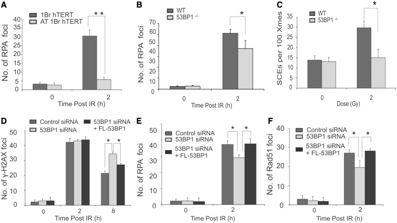
Figure 2.
Analysis of HR repair in immortalized human fibroblasts, in mouse embryonic fibroblasts and in complemented tumour cells. (A) Immortalized WT and A-T (AT1BR hTERT) deficient human fibroblasts were exposed to 3 Gy IR, and RPA foci were enumerated at 2 h post-IR. G2 cells were identified by CENPF staining. Aphidicolin was added before IR to prevent S phase cells progressing into G2 during analysis in all experiments. (B–C) Enumeration of RPA foci (B) and SCEs (C) in Wild-type (WT) and 53BP1−/− MEFs following IR. The statistical significance was determined using Student t-test. Asterisks indicate P < 0.05, double asterisks indicate P < 0.001. (D–F) U2OS cells were transfected with an siRNA oligonucleotide for 48 h to deplete 53BP1. Cells were then transfected with a plasmid encompassing an siRNA resistant full-length (FL) 53BP1 cDNA. Twenty-four hours later, cells were irradiated and enumerated for γH2AX (D) RPA (E) or RAD51 (F) foci. Foci were scored in HA+ cells, a marker present on the plasmid, providing a marker for cells that had undergone transfection. Results represent the mean and s.d. of three experiments. The statistical significance was determined using Student t-test. Asterisks indicate P < 0.05.

