Figure 2.
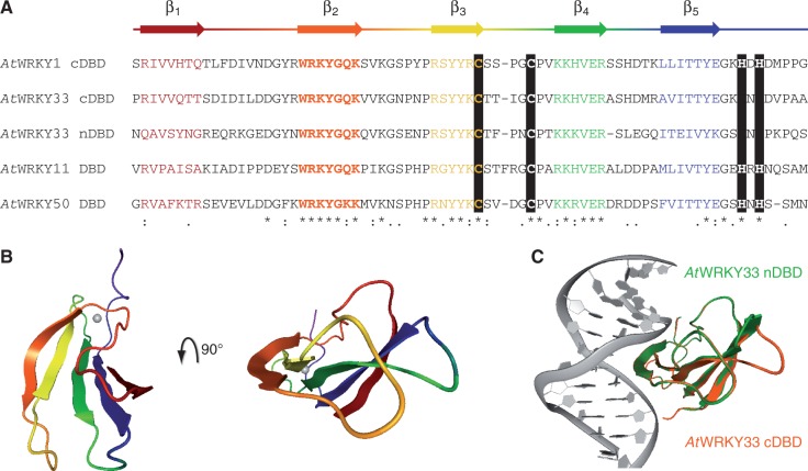
Homology models of AtWRKY DBDs. (A) The general protein secondary structure based on the crystal structure of WRKY1 C-terminal DNA-binding domain (cDBD; PDB id: 2ayd) is given above the alignment of AtWRKY cDBDs and N-terminal DBD (nDBD). Black bars highlight the conserved zinc finger; other conserved amino acids are indicated: (*) same amino acid, (:) amino acid with similar chemical properties, (.) majority of amino acids with similar chemical properties. (B) The five conserved β-strands of AtWRKY1 cDBD are colored according to A in the structure shown (PDB id: 2ayd). (C) The overlay of the protein-DNA models of AtWRKY33 cDBD (orange) and WRKY33 nDBD (green) is displayed. The protein structures are homology models and superimposed with respect to their β-sheet Cα atoms.

