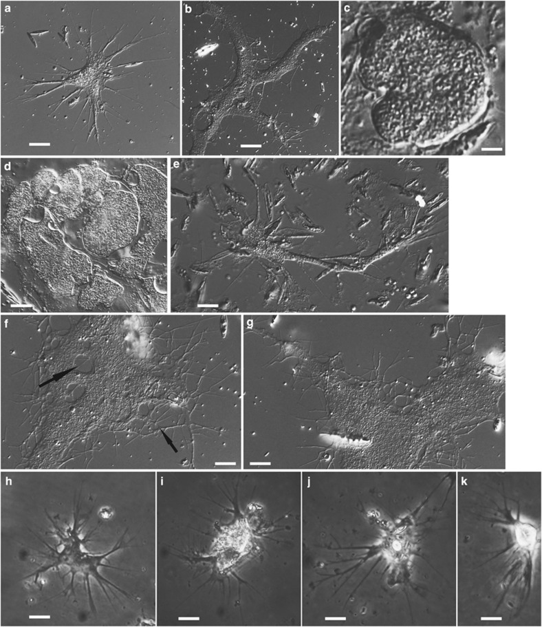
Figure 3.
Differential interference contrast light micrographs of isolate MVa1x (Thalassomyxa sp., Majorca, Spain, marine sediment) and phase contrast light micrographs of isolate NVam1 (Penardia sp., North Carolina, USA, brackish sediment). (a–g) Thalassomyxa sp. from Majorca; six different individuals (f and g are the same). Younger, more rapidly moving cells have a relatively compact (a) but sometimes elongated (e) morphology, similar to that of isolates KibAr and V1ld4 (see Figure 2). They can fuse into very large, slowly moving plasmodia (b, f, g) with lacunae in the cytoplasm and anastomoses between the peripheral filopodia (highlighted with arrows in f). Both smaller cells and giant plasmodia go into digestive cysts at regular intervals (c, d); a single plasmode can break up into many cysts of variable shapes and sizes (d). (h–k) Isolate NVam1 (Penardia sp.; four different individuals). Cell body generally intermediate between the more compact morphotype of some marine isolates and the branched morphotype of the terrestrial ones, but on average smaller than in all other isolates. Filopodia emerge from all around the cell; they are usually broader at the base and branching into thinner extensions. Temporary anastomoses frequently observed at the base of filopodia. Scale bars: 30 μm in a, e–g; 25 μm in b; 20 μm in d; 10 μm in h, i; 5 μm in c, j, k.

