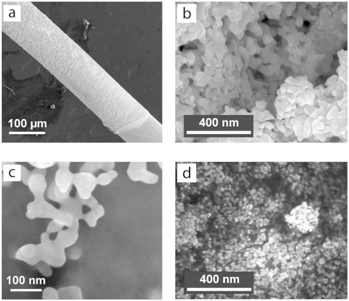
Figure 3. Scanning electron microscopy images of AuNPs modified microelectrodes.

(a) Low magnification image of the AuNPs modified Au microwire: the microwire geometry and the AuNP coating border can be seen in the lower right corner. (b) High magnification SEM image in the middle of the electrode in panel a. (c) High magnification SEM image of the border of the electrode in panel a. The images in panels a, b, and c were taken after the electrode modification with AuNPs and treatment in 0.5 M H2SO4. (d) SEM image of a reference sample covered with AuNPs identical to those used for the construction of nanobiostructures, the original nanoparticle shape can be clearly seen.
