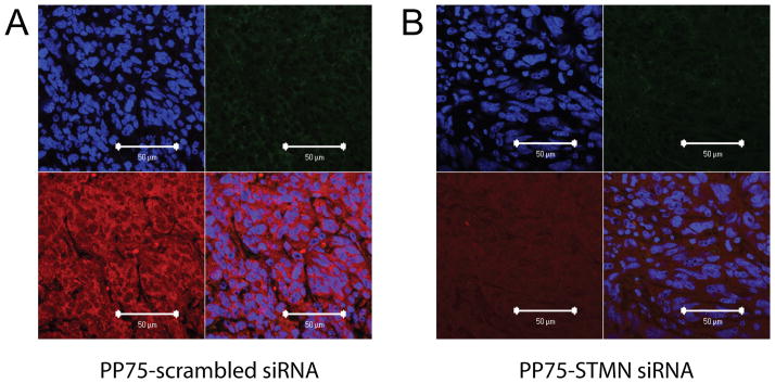
Fig 6. Assessment of immune infiltrates.

Subcutaneous flank tumors injected withPP75-scr siRNA (A) or PP75-STMN siRNA (B) conjugates were harvested three days later and processed for immunohistochemical analyses. Tissue sections were counterstained with DAPI to reveal cell nuclei (upper left panels). The presence of immune cell infiltrates was assessed by staining with an anti-CD11b primary antibody followed by a green fluorescently labeled secondary antibody (upper right panels). Stathmin expression was assessed using an anti-stathmin primary antibody followed by a red fluorescently labeled secondary antibody (lower left panels). A merged photomicrograph of the upper left, upper right and lower left panels is shown in the lower right panels. The scale bar in the lower right corner of each panel represents 50 μm.
