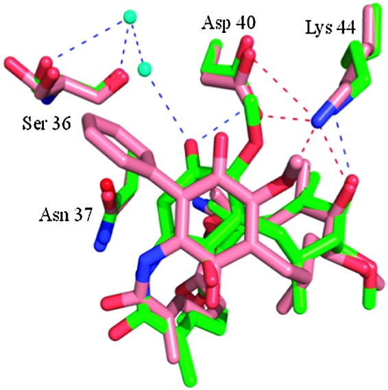
Fig. 2. X-ray structure of 19-phenyl geldanamycin 5 bound in the ATP site of yeast Hsp90.11 Geldanamycin 1 (green) and 19-phenyl geldanamycin 5 (salmon) with Hsp90 (green and salmon residues, respectively). See PDB codes ; 1A4H (geldanamycin 1) and ; 4ASF (19-phenyl geldanamycin 5).