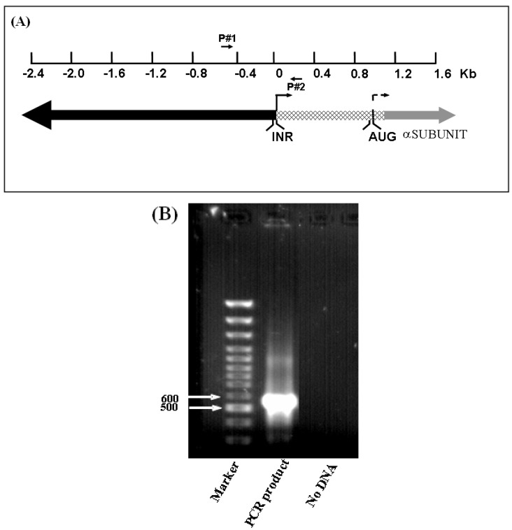
Figure 1.
IGF-IR promoter. (A) Schematic representation of the IGF-IR promoter region [5]. The initiator (INR) element is denoted by an arrow. The coding region, starting with the AUG codon, is shown in gray. The translation start site is denoted by a dashed arrow. The 5’-flanking region is denoted by a black bar, and the 5’-untranslated region (UTR) is represented by a dotted bar. The location of primers (P#1 and P#2), employed to amplify the proximal promoter is indicated. (B) PCR amplification of the human IGF-IR promoter. The PCR reaction was performed using a biotinylated antisense primer, as described under Materials and Methods section, and 4 μL of the biotinylated PCR product was loaded on a 1% agarose gel. A PCR reaction without DNA served as negative control. A 100-bp DNA ladder was used as a M.W. marker.

