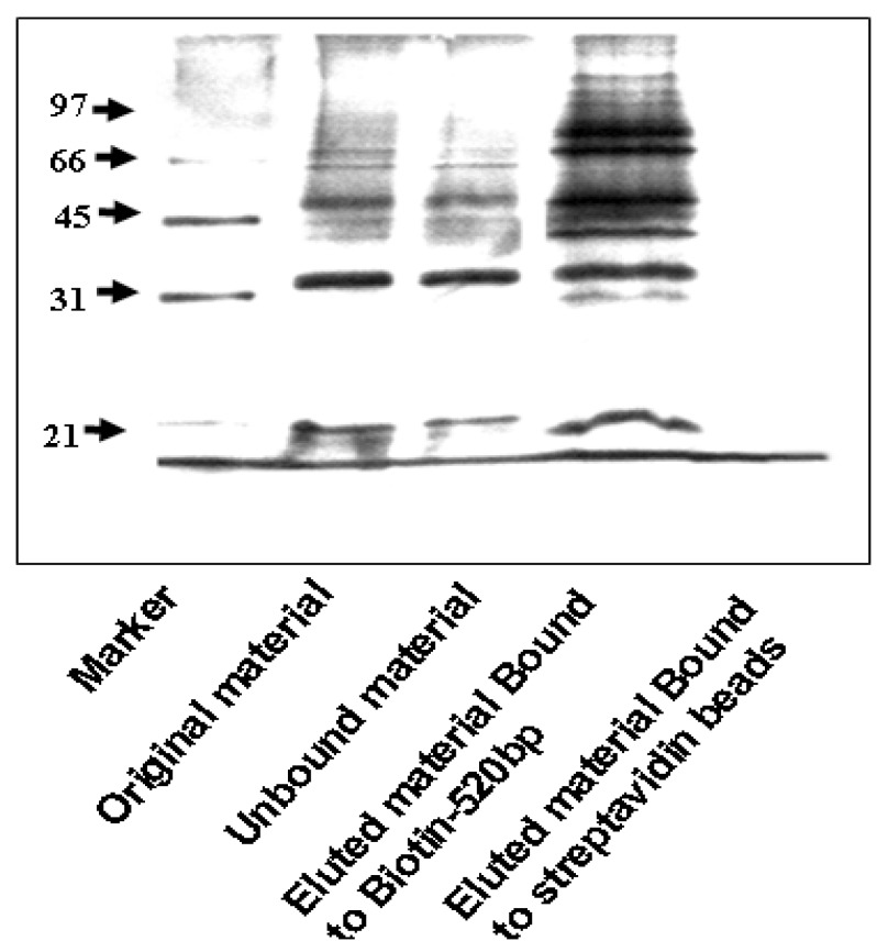
Figure 2.
Silver staining of IGF-IR promoter-bound proteins identified by DNA affinity chromatography. Nuclear extracts of MCF7 cells were prepared as described under Materials and Methods Section. Proteins bound to IGF-IR promoter DNA (10 μg) were electrophoresed through 10% SDS-PAGE, fixed, and stained with silver (Bio-Rad, Hercules, CA, USA). Lane 1, M.W. marker; lane 2, starting material (MCF7 nuclear extract, 4.2 μg); lane 3, unbound material (MCF7 nuclear extract that did not bind to DNA, 4.2 μg); lane 4, eluted material bound to Biotin-520 bp (MCF7 nuclear proteins that bound to IGF-IR promoter, 10 μg); and lane 5, negative control (MCF7 nuclear proteins bound to strepavidin magnetic beads, 10 μg).

