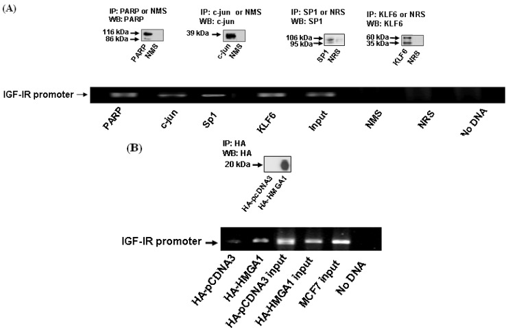
Figure 6.
ChIP assays of transcription factor binding to IGF-IR promoter DNA. (A). MCF7 cells were cross-linked with formaldehyde, lysed, sonicated, and immunoprecipitated with PARP, c-jun, Sp1, or KLF6 antibodies, or normal mouse or rabbit sera, followed by PCR amplification of precipitated chromatin using primers encompassing the IGF-IR promoter. The position of the 773 bp-amplified fragments is indicated. The input bands represent the amplified PCR product in the absence of antibodies. The insets indicate the endogenous levels of expression of the various proteins as measured by Western blots (WB) with specific antibodies or NRS or NMS as negative controls. IP, immunoprecipitation. (B). MCF7 cells were transfected with an expression vector encoding HA-HMGA1 (or empty pcDNA3 vector), after which the cells were cross linked with formaldehyde, lysed, sonicated and immunoprecipitated with an HA antibody, followed by PCR amplification of IGF-IR promoter DNA. HA-HMGA1 immunoprecipitated protein was detected by Western blots using an anti-HA antibody (inset).

