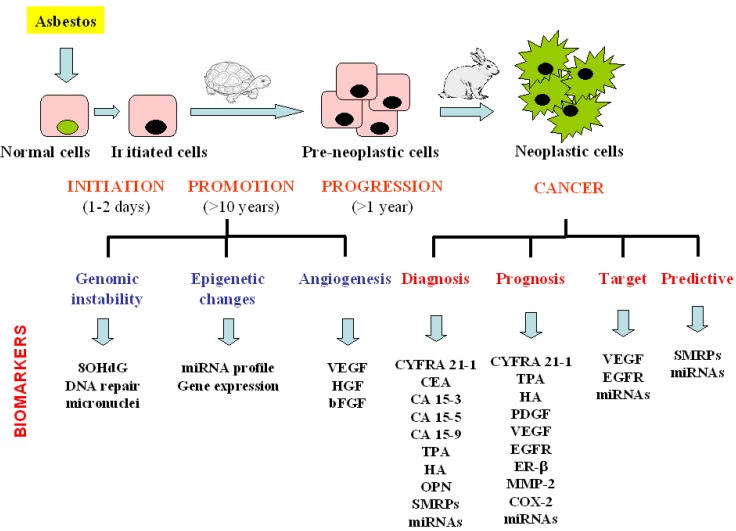
Figure 3.
Schematic presentation of biomarkers evaluated from asbestos exposure to malignant mesothelioma development. MM is characterised by a long latency period from the time of exposure to clinical diagnosis. The biomarkers that can be detected at the different phase of the malignant disease development are summarised.
8-hydroxy-2’-deoxyguanosine (8OHdG), vascular endothelial growth factor (VEGF), hepatocyte growth factor (HGF), basic fibroblast growth factor (bFGF), cytokeratin fragment (CYFRA 21-1), carcinoembryonic antigen (CEA), carbohydrate antigen 15-3 (CA 15-3), carbohydrate antigen 15-5 (CA 15-5), carbohydrate antigen 15-9 (CA 15-9), tissue polypeptid antigen (TPA), hyaluronic acid (HA), osteopontin (OPN), soluble mesothelin-related peptides (SMRPs), megakaryocyte potentiating factor (MPF), platelet derivate growth factor (PDGF), epidermal growth factor receptor (EGFR), matrix metalloproteinases-2 (MMP-2), cyclo-ocygenase-2 (COX-2).

