
Fig. 1.
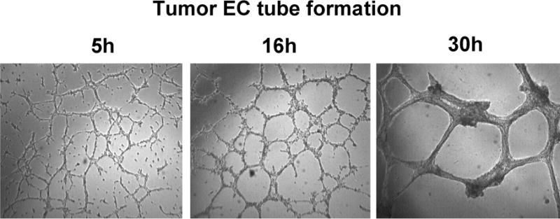
Time course of tumor endothelial cell tube formation.
Tumor endothelial cells (3B11, 2× 10⁵/ml) were incubated with matrigel and the tubes were imaged at 5, 16, and 30h, respectively, under an inverted phase microscope with a Leica DC350F CCD camera at 5× objective magnification using computer-controlled ProMax software.