
Figure 3. Incorporation of 40 kD protein in the sarcomere.
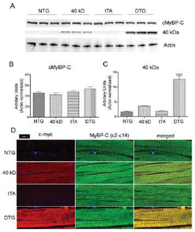
A, Myofibrils from NTG, 40 kD and tTA single TG and DTG mice expressing the 40 kD fragment were isolated, electrophoresed on acrylamide and subsequently transferred for Western blot analyses with cMyBP-C antibody. Actin was used as a loading control. B, Quantification of total, full length MyBP-C. No significant differences were found between the NTG, 40 kD, tTA and DTG groups. C, Quantification of 40 kD protein expression. Values represent mean±SE for each group (n=3). ***P<0.0001 DTG versus NTG, 40 kD, tTA controls. D, Incorporation of myc-tagged 40 kD cMyBP-C was confirmed in NTG, tTA, 40 kD and DTG mice by immunofluorescent staining of cMyBP-C with either an anti-myc (red) or anti cMyBP-C antibody (green).