
Figure 6. Activation of MEK-ERK signaling pathways in hearts expressing the 40 kD fragment.
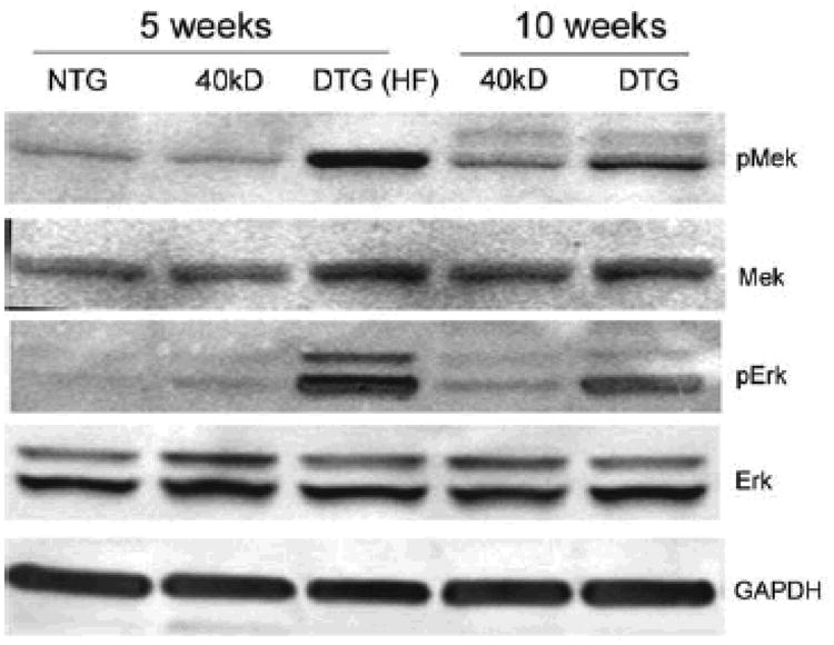
Heart lysates from 5 and 10 week old NTG, 40 kD single and DTG mice were examined for pMEK and pERK by Western blotting. The MEK-ERK pathways are highly activated in the DTG hearts, when compared with the NTG and single TG mice.