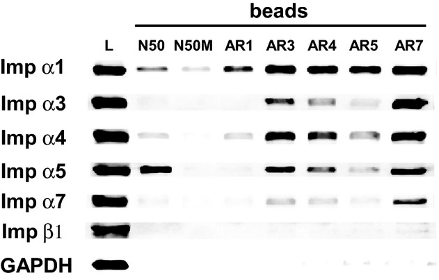
Figure 7.

Interaction of endogenous importins in Jurkat T cells with peptides derived from their auto‐inhibitory regions. Biotinylated N50 and N50M peptides, as well as biotinylated peptides representing the auto‐inhibitory regions (AR) of Imp α1, α3, α4, α5, and α7, were added individually to whole cell lysates from Jurkat T cells. The peptide and its interacting proteins were pulled down with SA‐coated beads and analyzed by immunoblotting with antibodies against a panel of importins as in Figure 3. Immunoblotting for the cellular protein GAPDH was used as a control to detect non‐specific interactions. Detection of endogenous proteins in the whole cell lysate is shown in the first column (lysate). Imp α indicates importin α; SA, streptavidin.
