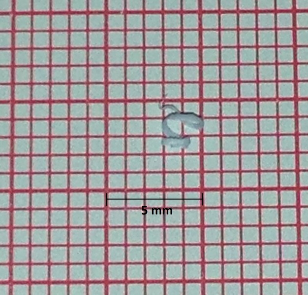
Figure 3.

Pictured is an example of grossly visible particles that were produced after the introduction of a conventional needle through the dilator and long sheath. The particles are placed on conventional electrocardiography paper as a size reference.
