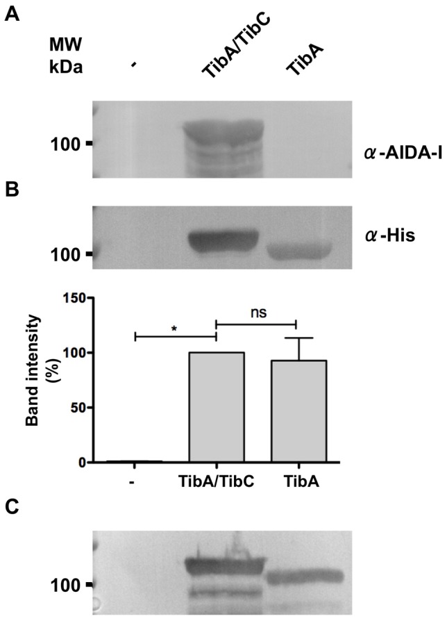
Figure 1. Effect of glycosylation on the expression level of TibA.

Whole cell extracts of E. coli strain C600 bearing an empty vector (-), or a plasmid allowing the expression of glycosylated TibA (TibA/TibC) or unglycosylated TibA (TibA) were separated by SDS-PAGE and revealed by immunoblotting with an anti-AIDA-I antibody (A) or an anti-His antibody (B). The intensity of the bands probed with the α-His antibody was quantified using ImageJ and values were normalized to the amount of glycosylated TibA (lower panel). Experiments were done five times and ANOVA and Dunnett post-tests were used to identify significant (*; p<0.05) and non-significant (ns) differences with glycosylated TibA. (C) Outer membrane extracts of E. coli strain C600 bearing an empty vector (-), or a plasmid allowing the expression of glycosylated TibA (TibA/TibC) or unglycosylated TibA (TibA) were revealed by immunoblotting using the anti-His antibody.
