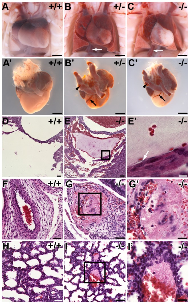
Figure 5. Pathological analysis of non-surviving Pdlim7 mutant pups reveals pre-mortem thrombi.

Pdlim7 +/- (n=3) and Pdlim7 -/- (n=11) perinatal lethal pups exhibit extensive blood clots in the heart, arteries (arrowhead), and veins (arrows, B-C’) associated with atrial dilation and lung congestion compared to WT (n=7) controls (A-A’). Example of pre-mortem blood clots in the right ventricle (E-E’), umbilical vessel (G-G’), and lung alveoli (I-I’) of a Pdlim7-/- perinatal lethal pup compared to WT control (D, F, H). Boxes depict location of high magnification image in E’, G’, and I’. Arrows point to attachment of the thrombus to the vessel wall (E’, I’) and arrowhead points to recanalization of the clot (G’). Scale bar = 1mm (A-C’), 50 µm (D-I), 20 µm (G’, I’), and 10µm (E’).
