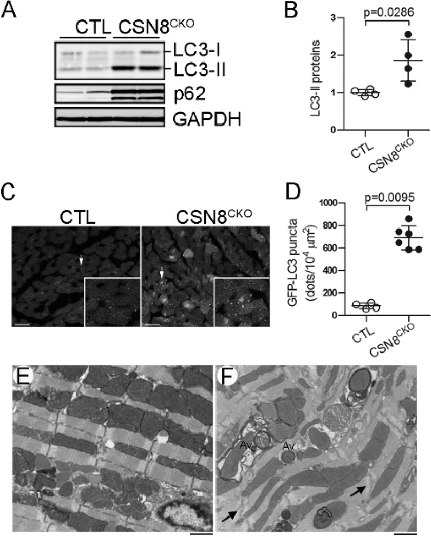
Figure 3. Marked increases of autophagic vacuoles in CSN8CKO hearts.
(A, B) Representative images of western blot analyses of LC3 and p62 (A) and a summary of LC3-II densitometry data (B) in CTL and CSN8CKO hearts at 5 days after the first tamoxifen injection. (C, D) Probing autophagy in CSN8CKO hearts using GFP-LC3. GFP-LC3 was introduced into the CTL and CSN8CKO mice via cross-breeding. Perfusion-fixed ventricular myocardium from CTL::GFP-LC3 mice and CSN8KO::GFP-LC3 littermate mice at 5 days after the first injection of tamoxifen was subjected to GFP-LC3 direct fluorescence confocal microscopy. Images from a 2.1 µm-thick slide of tissue were projected (C, representative images) and analyzed for the GFP-LC3 puncta density (D). The inset in panel C shows the area indicated by the arrow in a higher magnification. Bar=10 µm. (E, F) Electron micrographs of myocardium from CTL (E) and CSN8CKO (F) hearts. Abundant autophagosomes (Av) containing degenerating mitochondria and/or other cytoplasmic contents are evident in CSN8CKO hearts.

