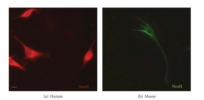
Figure 3.
Neurons detected by using anti-NeuH in immunofluorescence assays. In immunofluorescence assays, neurons in primary cell cultures of either human central nervous system (CNS) (a) or (b) mouse brain tissue were identified with the neuron-specific cellular marker, NeuH. The secondary antibodies used contained rhodamine (a) or FITC (b) as fluorochromes. In green, FITC signal in mouse neurons is shown and in red rhodamine signal is observed in human neurons. Original magnification was 400x, but the pictures were cropped to improve presentation.

