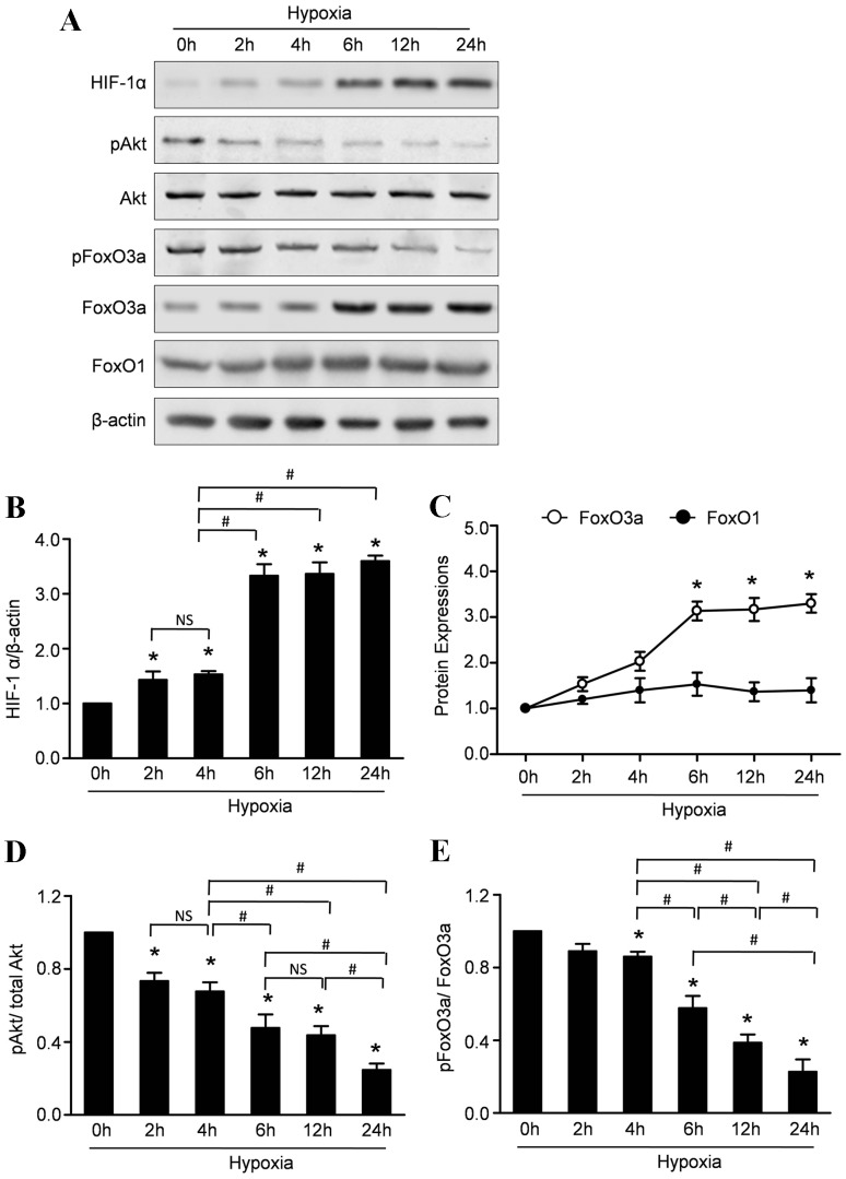
Figure 3. Hypoxia increases the expressions of HIF-1α, decreased the activity of Akt and regulates the expressions of FoxOs in CMECs.
A: Representative Western blots of HIF-1α, pAkt, Akt, FoxO1, FoxO3a, pFoxO3a in CMECs subjected to hypoxic injury. B: Semiquantitative analysis of HIF-1α. C: Semiquantitative analysis of FoxO1 and FoxO3a at the indicated time point. D: The ratios of p-Akt/total Akt at the indicated time point. E: The ratios of p-Akt/total Akt at the indicated time point. (n = 5,*p<0.05 vs. control, # p<0.05, NS: not significant).

