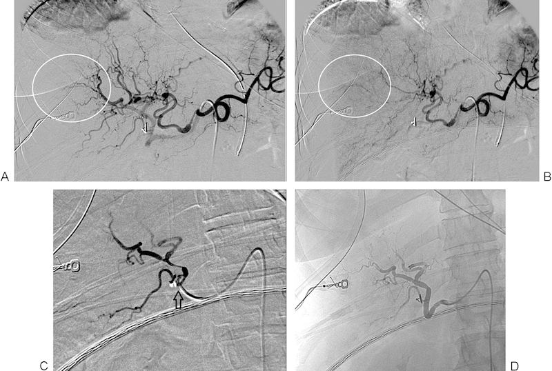
Figure 4.

(A) Digital subtraction angiogram of the celiac axis showing active extravasation of contrast from peripheral branches of the right hepatic artery (encircled). (B) Delayed image from the right hepatic arteriogram shows persistence of the extravasated contrast (encircled). (C) Postembolization right hepatic arteriogram shows pruning of the peripheral vessel branches and no further contrast extravasation. The catheter position (arrow) shows the point at which the embolization was performed. (D) Unsubtracted image from postembolization right hepatic arteriogram redemonstrating pruning of the peripheral vessel branches and no further contrast extravasation.
