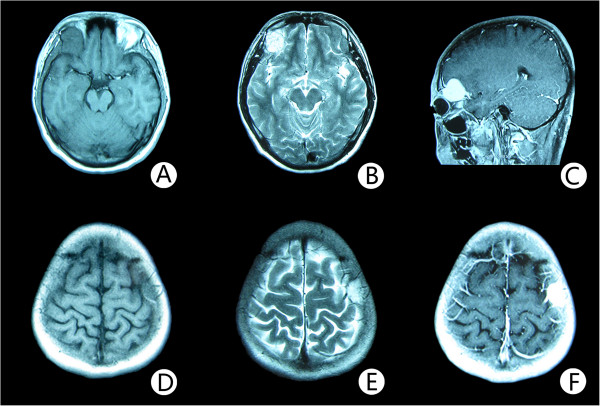
Figure 2.
Magnetic resonance image features of the two lesions. Noncontrast axial T1-weighted MR image (A) reveals a well circumscribed mass, measuring 3 cm × 3 cm × 3 cm, in the upward wall of the right orbit with intracranial extension. The lesion is well defined, and the signal intensity is of mixed intensity with some central hyperintensity. T2-weighted MR image (B) shows a lesion of high signal intensity. There is no surrounding edema. After administration of gadopentetate dimeglumine (C), strong and homogeneous enhancement of the mass and a dural tail sign is observed. The lesion extended intracranially and appears to compress the underlying brain. The MR image of the mass in the frontal bone over the central sulcus (D; E; F) is similar to the one in the upward wall of the right orbit (A; B; C).

