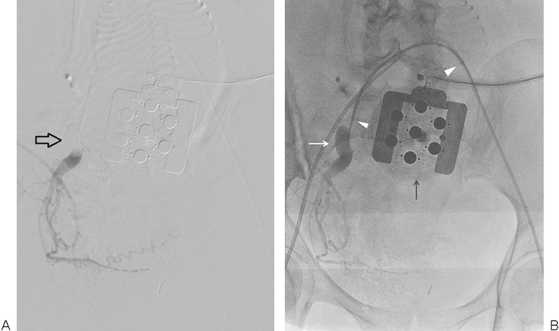
Figure 4.

A 34-year-old woman (G3P2) with complete placenta previa and percreta who underwent preoperative balloon catheter placement for scheduled cesarean delivery. (A) Right-sided hypogastric artery digital subtraction angiography (DSA) after balloon catheter inflation demonstrates good position of the occlusion balloon (open arrow) and stasis of forward flow. (B) DSA native image redemonstrates balloon catheter (white arrow) and bilateral 7-French sheath tips (arrowheads). Radio-opaque fetal monitoring equipment overlies the anterior abdomen (black arrow).
