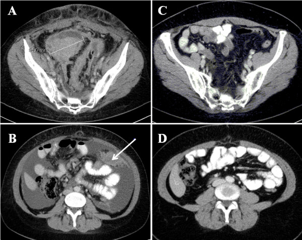
Figure 1.

Axial CT slices of patient # 3 after contrast material was intravenously administered. A target lesion with a SLD of 84 mm is localized near the minor pelvis before (A) and after (C) six cycles of first-line chemotherapy. (B) The arrow represents a mesenteric peritoneal carcinosis (non-target lesion) in the same patient that is level with the lower edge of the liver prior to chemotherapy. A significant amount of ascites associated with the non-target lesion is also observed (B). (D) Both non-target lesions are absent after the completion of chemotherapy.
