
Figure 5.
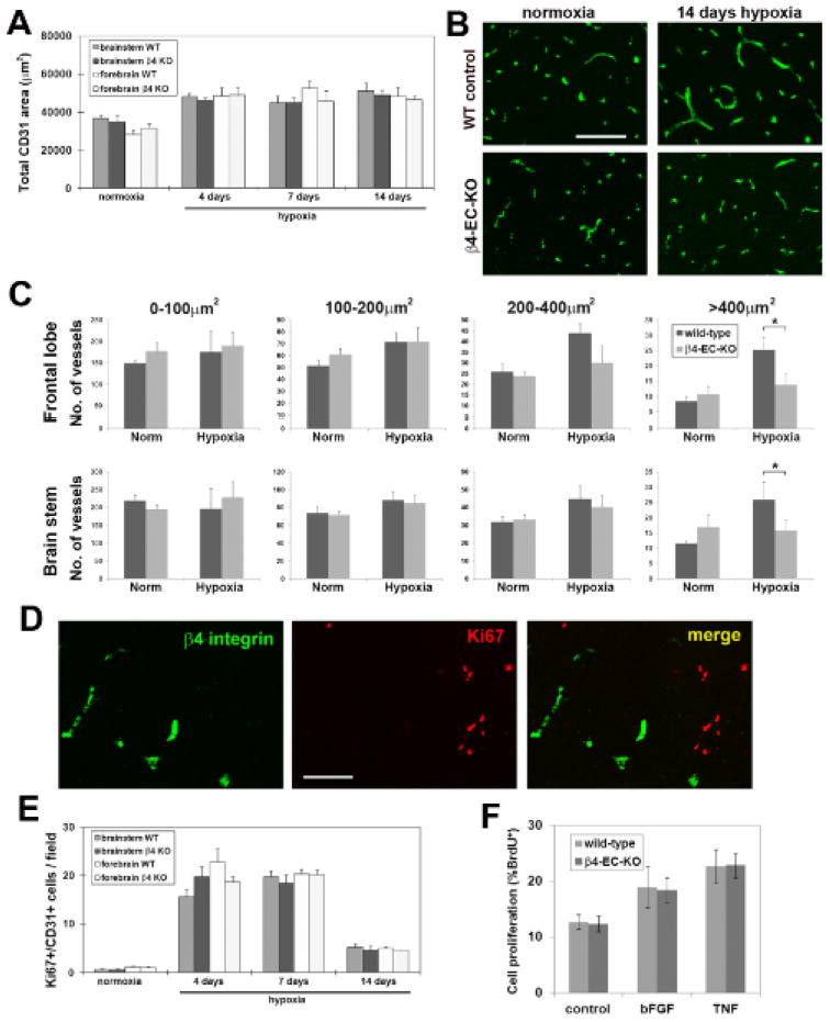
Comparison of hypoxic-induced vascular remodeling in the brains of wild-type and β4-EC-KO mice. β4-EC-KO and wild-type mice were maintained at normoxia or exposed to mild hypoxia (8% O2) for 4, 7 or 14 days before frozen brain sections were immunostained to determine the influence of hypoxia on: A. total vascular area, B and C. cerebral vessel size distribution, and E. endothelial cell proliferation. Analysis was performed with four different animals per condition, and the results expressed as the mean ± SEM. Note that cerebral hypoxia promoted similar increases in total CD31 area (A), and in the number of proliferating BEC in the brains of wild-type and β4-EC-KO mice (E), with no significant differences observed between the two groups at any time-point. However, vessel size distribution revealed an important difference between wild-type and β4-EC-KO mice (C). In wild-type mice, 14 days hypoxia induced a preferential increase in the number of vessels in the size range of arterioles (area > 400μm2) in both brain areas examined. In contrast, β4-EC-KO mice failed to show this response. * P< 0.05. D. β4 integrin/Ki67 dual-IF of 7 day hypoxic wild-type brain. Scale bars in panels B and D = 100μm. Note that despite the presence of many Ki67-positive cells in the hypoxic remodeling brain, dual-labeled β4 integrin/Ki67 cells were never detected. F. Comparison of cell proliferation by BrdU incorporation in BEC derived from β4-EC-KO or littermate control mice. Note that while bFGF and TNF increased BEC proliferation relative to control conditions, no differences were observed in the mitotic rates of BEC derived from β4-EC-KO or littermate control mice.