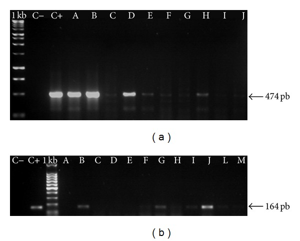
Figure 2.

Example of BPV DNA sequences detected in cutaneous lesions and in respective primary culture cells: the samples were collected from one of the animals. I primers FAP59/64, II BPV2 specific primers. C− negative control, C+ positive control, A-B PCR with DNA from papilloma lesion, C–E primary cell culture using fragments from BPV positive lesion in passage 1, F–H primary cell culture using fragments from BPV positive lesion in passage 2, I-J primary cell culture using fragments from BPV positive lesion in passage 3, L primary cell culture using fragments from BPV positive lesion in passage 4, and M primary cell culture using fragments from BPV positive lesion in passage 5. It is important to pay attention in the different amplicons, suggesting possible different virus load.
