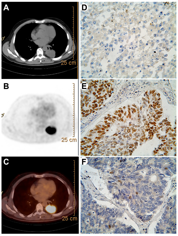
Figure 1.
Representative images of PET-CT and immunohistochemistry. Transaxial images of (A) diagnostic CT, (B) FDG-PET and (C) fusion of PET and CT images. Immunohistochemical stainings for (D) epidermal growth factor receptor (EGFR), (E) p53 and (F) excision repair cross complementing gene 1(ERCC1). (magnification, ×400).

