Fig. 1.
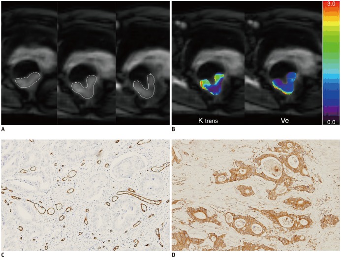
Dynamic contrast-enhanced magnetic resonance imaging image of pathologically T2N0 rectal cancer from 50-year-old woman.
Three serial ROIs were drawn from three sections through tumor (A), and mean Ktrans (1.174 min-1) and Ve (0.209) were obtained (B). Histopathologic specimen of rectal adenocarcinoma showed (C) high MVD score (vascular endothelial cells shown in brown identify microvessels) and (D) strong VEGF expression (positive expression of VEGF is shown in brown in cytoplasm; 100 ×). Hot-spot MVD was 99.3 (mean number of microvessel/mm2), and VEGF expression score was 7. ROI = region of interest, MVD = microvessel density, VEGF = vascular endothelial growth factor

