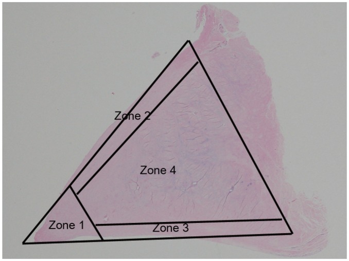
Fig. 1.

Meniscal zones. Meniscus is divided into four zones, including apical surface (zone 1), superior lamellar layer (zone 2), inferior lamellar layer (zone 3), and central layer (zone 4). Apical surface (zone 1) is defined as area occupying approximately 20% of inner diameter of meniscus on slide. Superior and inferior lamellar layers are composed of superficial network (about 10 micrometers thick) and lamellar layer (about 150-200 micrometers thick). Four zones of meniscus are drawn on histologic photomicrograph (hematoxylin & eosin staining, 1 : 1) of normal meniscus.
