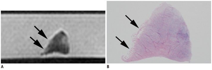
Fig. 3.

Seventy-eight-year-old female with minimal tear in superior lamellar layer.
A. MR image of meniscal section shows high signal intensity (S1) and surface irregularity (M1) (black arrows) in superior lamellar layer. B. Histologic photomicrograph (hematoxylin & eosin staining, 1 : 1) shows minimal tear in superior lamellar layer and apical surface (black arrows).
