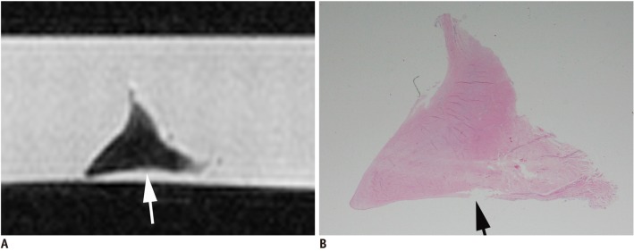
Fig. 4.

Seventy-eight-year-old male with meniscal thinning.
A. MR imaging of medial meniscus shows surface irregularity on inferior lamellar layer (white arrow). B. Photomicrograph (hematoxylin & eosin staining, 1 : 1) shows thinning of inferior lamellar layer (black arrow).
