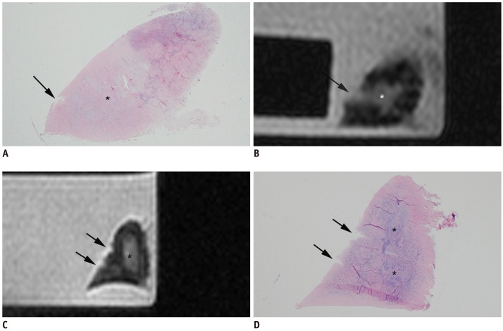
Fig. 6.
Sixty-eight-year-old female with MR Grade 3 signal intensity (A, B), and 60-year-old female with minimal tear of superior lamellar layer, which is distant from degeneration of central layer (C, D).
A. On histologic photomicrograph (hematoxylin & eosin staining, 1 : 1), minimal tear of superior lamellar layer (black arrow) is connected to degeneration of central layer (asterisk). B. MR image of same section shows MR grade 3 signal intensity (black arrow), in which abnormally high signal intensity of minimal tear is connected to high signal intensity of adjacent central layer of meniscus (asterisk). C, D. MR image shows high signal intensity and surface irregularity due to minimal tear (black arrows) of superior lamellar layer, which is confirmed on histologic slide D (hematoxylin & eosin staining, 1 : 1). Ovoid-shaped high signal intensity of central layer on MR image (asterisk in C), which is distant from minimal tear of superior lamellar layer, is shown to be degeneration of central layer on histological examination (asterisks in D). MR finding of this meniscal specimen was considered to be of grade 2 signal intensity.

