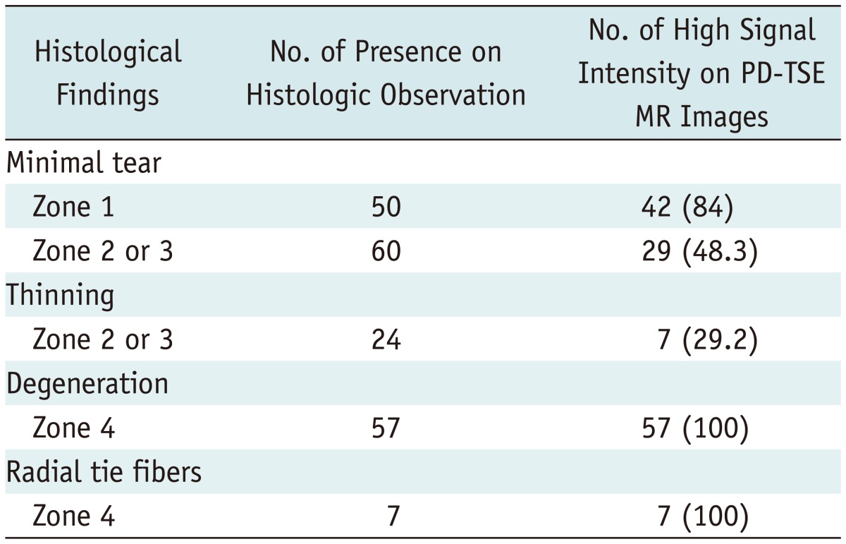
Table 3.
Summary of Histological and MR Findings of Meniscal Specimen

Note.- Total of 100 meniscal specimens were examined. Meniscus is divided into four zones including apical surface (zone 1), superior lamellar layer (zone 2), inferior lamellar layer (zone 3), and central layer (zone 4). Data in parentheses are percentages of observations. PD-TSE = proton density-weighted turbo spin-echo
