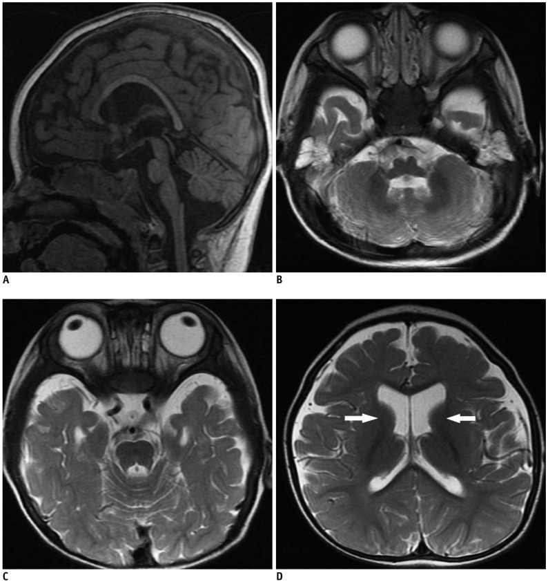
Fig. 1.
MR images of 1 year and 4 months girl with Cri-du-Chat syndrome.
Sagittal T1-weighted (A) and axial T2-weighted (B-D) images show hypoplasia of brainstem, most prominently in pons, with normal cerebellum, thinning of corpus callosum and mega cisterna magna (A, C). Mild atrophy of both frontal and temporal lobes and decreased myelination in both anterior limbs of internal capsules (arrows) are seen (D).

