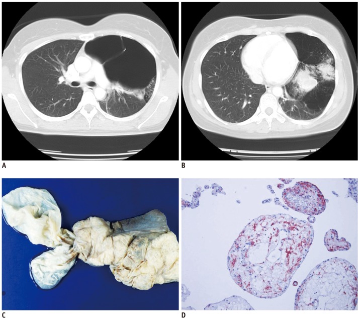
Fig. 1.
Thirty-one-year-old female patient with large bullae and subsegmental consolidation in left upper lobe.
A. Preoperative chest computed tomography shows multiple giant bullae in left upper lobe with resultant mediastinal shifting. B. Subsegmental consolidation in left lingular segment has broad contact with bullae. C. Pleural surface shows several massively dilated bullae, largest one measuring 11 × 5 × 3 cm in size. D. On high power, villous structures are composed of simple cuboidal cell linings and edematous cores. Immunohistochemical stain for CD-10 is positive only in interstitial cells (IHC, × 400).

