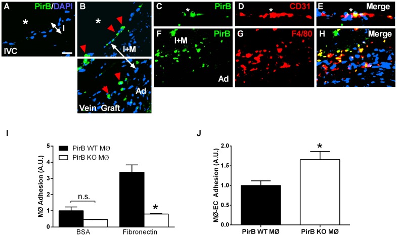
Figure 2. PirB mediates adhesion in vitro and in vivo.
(A) Representative immunofluorescence image showing lack of PirB signal in the pre-implantation vein. IVC, inferior vena cava. Scale bar, 20 µm. (B) PirB positive cells localized to the luminal surface and the interface between the medial and adventitial layers. *, vessel lumen; red arrowhead, PirB positive cells; I, intimal layer; I+M, intima-medial layer; Ad, adventitial layer. (C–E) PirB-positive cells on the vein graft luminal vessel surface (C) did not co-localize with CD31-positive endothelial cells (E). *, vessel lumen; white arrows, PirB-positive cells. (F–H) PirB-positive cells in between the medial and adventitial layers (F) co-localize with F4/80-positive cells (G,H). I+M, intima-medial layer; Ad, adventitial layer. n = 4. (I) Bar graph shows macrophage adhesion to bovine serum albumin (BSA) or fibronectin. Macrophages were derived from PirB WT (▪) or PirB KO (□) mice. n.s., not significant. n = 8. *, p<0.0001; ANOVA; post-hoc testing p<0.05. (J) Bar graph shows macrophage adhesion to endothelial cells. Macrophages were derived from PirB WT (▪) or PirB KO (□) mice and activated with TNF-α. *, p = 0.0499, t-test; n = 3.

