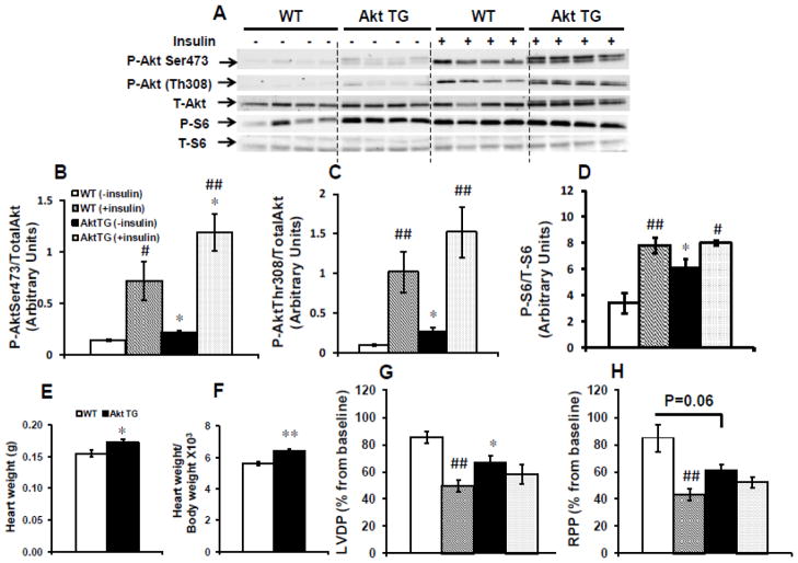
Fig. 5.
Akt activation in the heart abolished cardioprotection by IPC. Hearts isolated from Akt TG and WT mice were subjected to the full IPC protocol as shown in Fig. 1A in the presence or absence of 1 nmol/L insulin. (A) Representative western-blots of Akt phosphorylation on Ser473 and Thr308, total Akt, S6 phosphorylation on Ser235/236 and total S6 protein; (B), (C) and (D) the corresponding densitometry of phosphoSer473Akt/total Akt, phosphoThr308Akt/total Akt and phosphoSer235/236 S6/total S6 ratios respectively. (E) heart weights; (F) heart weight/body weight ratios ×103; (G) % recovery of left ventricular developed pressure (LVDP); (H) % recovery of rate pressure product (RPP) in Akt TG and WT mice. Data are mean ± SEM (4 mice per group and per genotype for western-blots and n = 5–8 mice per group and per genotype for functional recovery). *p<0.05; **p<0.005 versus WT perfused in the same condition; #p<0.05; ##p<0.005 versus (− insulin) within the same genotype.

