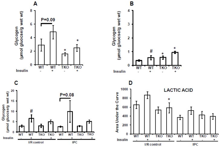
Fig. 7.
Glycogen and lactate levels in TIRKO and WT heats. (A) Pre-ischemic glycogen content in TIRKO and WT hearts subjected to 50 min perfusion without IPC in the absence or presence of 1 nmol/L insulin. (B) Same as (A) but hearts received three cycles (5 min each) of IPC. (C) Post-ischemic (at the end of the 45 min reperfusion) glycogen content in TIRKO and WT hearts subjected to I/R or IPC with or without 1 nmol/L insulin. (D) Lactate levels measured by GC-MS at the end the reperfusion in TIRKO and WT hearts subjected to I/R or IPC in the absence or presence of 1 nmol/L insulin. Data are mean ± SEM (n = 3–4 mice per group for A and B and n = 4–7 mice per group for C and D. *p<0.05 versus WT perfused in the same condition; #p<0.05 versus (− insulin) within the same genotype.

