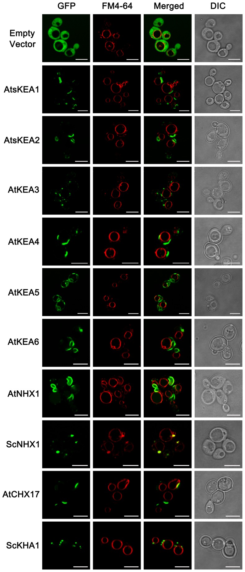
Figure 10. The diversified distribution of AtKEAs in yeast.

Wild-type (W303-1B) yeast strains harboring pDR196-GFP, pDR196-AtKEAs-GFP, pDR196-AtNHX1-GFP, pDR196-AtCHX17-GFP, pDR196-ScNHX1-GFP, and pDR196-ScKHA1-GFP (fused with GFP at the C terminus), respectively, were grown to logarithmic phase in SC-URA medium (pH 5.8) and were stained with FM4-64 dye. The subcellular localizations of the GFP-tagged proteins (green) and FM4-64 fluorescence (red) were observed under the Laser Scanning Confocal Microscope. Bars, 5µm.
